ByJohn A
When Cancer Affects Vision: Understanding the Neurological Connection
Vision problems are rarely the first symptom people associate with cancer, yet the connection between oncology and eye health runs deeper than most patients realize. Brain tumors, blood cancers, and metastatic disease can all manifest through visual disturbances, double vision, or sudden sight loss. For oncologists and ophthalmologists alike, these symptoms represent a diagnostic challenge that requires specialized expertise bridging neurology, vision science, and cancer care.
How Cancer Reaches the Visual System
The visual pathway extends from the retina through the optic nerves, optic chiasm, and into the brain’s occipital cortex. Any tumor or malignancy along this route can disrupt vision. Primary brain tumors such as gliomas or meningiomas may compress the optic nerve or visual cortex directly. Pituitary adenomas, though often benign, commonly press against the optic chiasm, causing characteristic peripheral vision loss in both eyes.
Metastatic cancers follow different patterns. Breast, lung, and melanoma cancers frequently spread to the brain, while leukemia and lymphoma can infiltrate the optic nerve itself or cause inflammation in the eye structures. Paraneoplastic syndromes—immune responses triggered by distant cancers—may attack the retina or optic nerve even when no tumor cells are present in the eye.
Warning Signs That Demand Urgent Assessment
Certain visual symptoms in cancer patients warrant immediate neurological and ophthalmological evaluation. Progressive vision loss in one or both eyes, especially when painless, suggests optic nerve involvement. Double vision that worsens when looking in specific directions points to cranial nerve damage from tumor pressure or infiltration. Sudden blindness in one eye could indicate optic nerve stroke, sometimes caused by tumor compression of blood vessels.
Peripheral vision defects deserve particular attention. A patient who bumps into doorframes or misses objects to one side may have hemianopia—loss of half the visual field—indicating damage to the brain’s visual processing areas. Papilledema, swelling of the optic nerve head visible during eye examination, signals increased intracranial pressure from brain masses. These findings require urgent imaging and often immediate intervention to prevent permanent vision loss.
The Diagnostic Process for Vision-Related Cancer Symptoms
Evaluating cancer-related vision problems requires coordination between multiple specialties. The assessment typically begins with detailed visual field testing to map exactly which portions of vision are affected. This pattern helps localize where along the visual pathway the problem originates. Optical coherence tomography provides high-resolution images of retinal layers, detecting subtle infiltration or swelling invisible during routine examination.
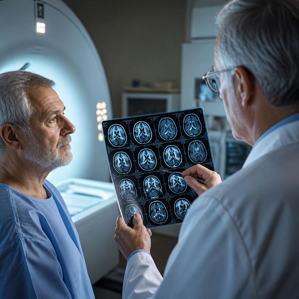
Brain imaging forms the cornerstone of diagnosis. MRI with gadolinium contrast reveals tumor masses, optic nerve enhancement suggesting inflammation or infiltration, and abnormal tissue in visual processing centers. PET-CT scanning helps distinguish active tumor from post-treatment changes, particularly important in patients with known cancer histories. Lumbar puncture may be necessary when leptomeningeal spread—cancer cells in the fluid surrounding the brain and spinal cord—is suspected.
Specialized Care for Complex Cases
Cancer patients experiencing neurological vision problems benefit from centers where oncology expertise intersects with advanced ophthalmological and neurological care. The multidisciplinary approach ensures that treatment plans address both the underlying malignancy and its visual consequences. Radiation oncologists, for instance, must carefully plan beam trajectories to treat tumors near the optic apparatus while minimizing radiation damage to these sensitive structures.
For patients in the Gulf region facing these complex diagnostic challenges, comprehensive cancer centers with dedicated neuro-ophthalmology programs offer the integrated expertise required. Neuro-ophthalmology Abu Dhabi services within major cancer institutes combine advanced imaging technology with subspecialty-trained physicians who understand both the oncological and visual dimensions of disease. This coordination becomes particularly crucial when treatment decisions must balance cancer control against preserving sight, or when visual symptoms represent the first clue to previously undetected malignancy.
Treatment Approaches and Visual Outcomes
Treatment strategies depend entirely on the cancer type, location, and extent of visual pathway involvement. Surgical resection offers the best chance for both cancer control and vision preservation when tumors are accessible and well-demarcated. Transsphenoidal surgery, performed through the nose to access pituitary tumors, can decompress the optic chiasm with minimal brain manipulation. However, infiltrative tumors wrapped around the optic nerve or invading the cavernous sinus present surgical risks that may outweigh potential benefits.
Radiation therapy has evolved substantially with techniques like stereotactic radiosurgery and intensity-modulated radiation therapy allowing precise targeting. These approaches deliver therapeutic doses to tumors while respecting dose limits for the optic nerves and chiasm, typically kept below 54 Gray to minimize radiation-induced vision loss. Fractionating treatment over multiple sessions further protects these structures while maintaining anti-tumor effectiveness.
Systemic treatments play expanding roles. Chemotherapy reaches tumors throughout the central nervous system, though the blood-brain barrier limits drug penetration. Newer immune-oncology and targeted therapies sometimes produce dramatic responses in previously treatment-resistant cancers. Lymphomas and leukemias involving the eye or optic nerve often respond well to chemotherapy, with vision recovery possible if treatment begins before irreversible damage occurs.
The Broader Healthcare Context
Managing cancer’s intersection with vision exemplifies broader challenges in modern oncology, where advancing technology and expanding treatment options have created both opportunities and complexities. The financial burden of comprehensive cancer care continues to grow as new diagnostics and therapies enter clinical practice. As detailed in analyses of strategies to manage cancer treatment costs, healthcare systems worldwide grapple with delivering cutting-edge care while containing expenses.
The integration of multiple specialties around complex cases like vision-threatening cancers represents one approach to improving both outcomes and efficiency. Rather than sequential consultations across disconnected providers, multidisciplinary tumor boards and integrated care teams reduce diagnostic delays and prevent conflicting treatment recommendations. This coordination becomes especially valuable when treatment decisions involve trade-offs between different goals—such as aggressive tumor control versus preserving quality of life through maintained vision.
Recovery and Long-Term Monitoring
Visual recovery following treatment varies widely. Some patients experience rapid improvement as tumor pressure on neural structures resolves. Others face permanent deficits from irreversible nerve damage that occurred before treatment began. Rehabilitation services, including vision therapy and adaptive strategies, help patients maximize remaining visual function. Low-vision specialists provide devices and training that enable continued independence despite partial sight loss.
Long-term surveillance remains essential. Regular visual field testing and optic nerve imaging detect recurrence or radiation-induced damage before symptoms appear. Patients treated with certain chemotherapy agents face ongoing risks of cataracts or retinal toxicity requiring ophthalmological monitoring. The connection between systemic health interventions and specialized outcomes appears in various medical contexts, similar to how emerging medications for metabolic conditions require careful monitoring to balance benefits against potential adverse effects.
Looking Forward
The intersection of oncology and neuro-ophthalmology will likely see continued innovation. Liquid biopsy techniques may soon detect tumor DNA in the aqueous humor of the eye, enabling less invasive monitoring of intraocular malignancies. Artificial intelligence algorithms already assist in detecting subtle optic nerve changes on imaging that human observers might miss. Gene therapies and next-generation immunotherapies may offer new options for cancers currently considered untreatable.
For patients and families confronting vision symptoms in the context of cancer, the message remains clear: these manifestations deserve prompt, specialized evaluation. The window for preserving vision can be narrow, making rapid access to integrated diagnostic services and treatment planning essential. As cancer care grows increasingly sophisticated, the collaboration between oncologists and vision specialists exemplifies how medicine’s various subspecialties must work together to address the full spectrum of disease impacts on human health and quality of life.